
05 August 2026
It takes skill to draw a cube from a square, a sphere from a circle, and most extraordinarily to draw 3D anatomy from a 2D image, in your head.
For Sonographers, learning this artform has long been the reality.
However, researchers at the Massachusetts Institute of Technology (better known as ‘MIT’) have envisioned a new reality - an augmented one - to help Sonographers bridge the skill gap and increase access to this already cost-effective and portable modality, all thanks to a headset, controllers and video game software.
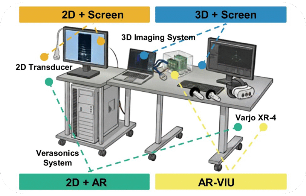
Combining ultrasound and Augment Reality (AR) with help from video game software, MIT's recent study – published online in nature.com – compared the accuracy of 9 'experienced' and 9 'novice' Ultrasound experts across imaging tasks between traditional visualisation methods and their new AR-VIU (Augmented reality, Real-time, Volumetric Imaging in ultrasound) system.
AR-VIU links the ultrasound probe with an AR controller. As the probe takes volumetric scans, the data is translated into a virtual 3D model that users can visualise, scale and manipulate in real space and real time through an AR headset.
The 3D model is made possible by sending the probe's data through to an external computer running 'Unreal Engine' - a video game rendering software - which translates the data into a full 3D model, running on a laptop with a dedicated Graphics Processing Unit (GPU).
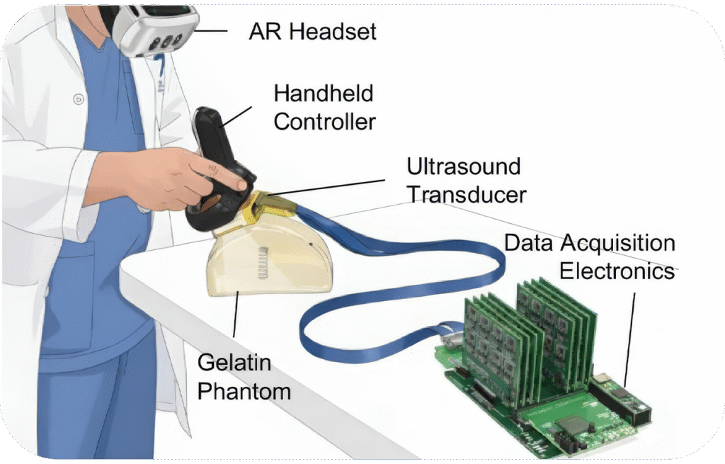
This allows practitioners to view the scanned anatomy as it exists in real space, while also manipulating a scaled up, detailed model for more precise diagnostics without the limitations of a 2D screen or Phantasia - the psychological ability to visualise objects in the mind.
Comparatively both groups recorded high identification accuracy when using the AR-VIU system, and overall when using 3D visualisation methods against conventional 2D methods.
While the ‘experienced’ group demonstrated high accuracy across all visualisation methods; the novice group, however, demonstrated a considerable skill gap - scoring a lower median accuracy of ~50% - while using the conventional 2D screen method.
Notably, as 3D imaging and AR technology were progressively included in each visualisation, the novice group began scoring increasingly comparable accuracy to the experienced group at a median accuracy close to 100% - successfully bridging the existing skill the gap between the groups.
The success of the AR-VIU system and others like it could become a vital catalyst in the training and development of Sonographers; in addition to widening its accessibility among multi-disciplinary radiographers such as those in rural and regional Australia.
"For training, this could make ultrasound more intuitive and more understandable." said senior author of the study and associate professor of media and art sciences at MIT, Canan Dagdeviren.
"On the clinical side, it could be less time consuming, more accurate, and also give more health care providers more piece of mind. They wouldn't have to wonder if they missed anything."
Augmented Reality equipment - like Virtual Reality - is compact and commercially available as are laptops with dedicated GPU's, meaning systems like AR-VIU could be readily integrated with existing, portable ultrasound machines.
Following the study, participants were surveyed on their preference of visualisation method.
While the novice group showed an overwhelming preference for AR-VIU, the experienced group showed a bell curve of moderate interest; instead recording their highest median preference for the conventional and familiar 2D screen method.
Like many MRP's, Sonographers are increasingly in demand, especially across rural and remote areas.
If successful, MIT's AR-VIU technology and others like it could provide invaluable training opportunities for new and existing Sonographers - alongside multidisciplinary radiographers - to meet the ever-growing demand for diagnostic imaging.







© ASMIRT 2026 | ALL RIGHTS RESERVED | PRIVACY | TERMS | SITEMAP